In the general care of the eyes, the only thing that the average dog owner can be advised to do is to keep them clean.
For safety’s sake, any injury to the dog eye should be considered an emergency and the veterinarian should be contacted immediately. Professional attention should be given even to very mild dog eye irritations that do not readily respond to simple treatment with washings of boric-acid solution or applications of boric-acid eye ointment.
Eye conditions are so complex that even the veterinarian occasionally has to solicit the assistance of a human eye specialist in order to arrive at a correct diagnosis and to determine a proper course of treatment. It is apparent, therefore, that a detailed discussion of eye diseases would be useless for the nonprofessional person. It would be well, however, to describe briefly a couple of the simpler eye ailments in order to give the reader at least a casual idea of what he may encounter.
Table of Contents
Conjunctivitis in dogs
The membrane that lines the eyelids and covers the front of the eyeball is called the conjunctiva. An inflammation of this membrane is called conjunctivitis. Where only the eye itself is involved, the condition is called a primary conjunctivitis. Where the eye irritation is accompanied by another disease, such as rabies or distemper, the condition is called a secondary conjunctivitis. Mild forms of primary conjunctivitis are usually not serious if given prompt attention. If the condition is neglected, however, it often becomes increasingly resistant to treatment.
Conjunctivitis may be caused by infection, injury, dust or chemical irritation, or, as indicated above, it may be a secondary symptom of other primary conditions such as rabies, distemper, and many other infectious or parasitic diseases. It also appears commonly as a secondary symptom of various types of stomach upsets, and usually is one of the first indications of any indisposition of the animal. That is why the veterinarian always checks the eyes during a routine examination.
Conjunctivitis is characterized by redness of the conjunctiva with an accompanying eye discharge that may vary from a watery consistency to a creamy accumulation of pus. The animal may rub the affected eye with its paws, and this mechanical irritation will cause further swelling and redness of the conjunctiva and the eyelids. There may be small granular elevations on the conjunctiva, the appearance of a false membrane, or occasionally deeper tissues may become involved. Resistant and neglected cases may become chronic and lead to more serious infections that may ultimately impair vision.
Conjunctivitis is treated mainly by the application of suitable antiseptics and healing agents, either in liquid or ointment form. Often the veterinarian will inject certain protein substances into the body to assist in the healing process. These substances cause an artificial increase in the production of white blood cells, and these white blood cells are the good soldiers of the blood that help to fight infection. In some cases, bacteriological examinations of the eye discharge may be made in order to identify the causative organisms, and then the specific drugs which can eradicate them may be applied. Such wonder drugs as penicillin, aureomycin and terramycin have proved very useful in these diseases. Where conjunctivitis is a secondary complication, the elimination of the primary condition usually results in the disappearance of the conjunctivitis.
Dislocated eyeball in dogs
In dogs, dislocation of the eyeball is an emergency. If the eyeball is not replaced within a couple of hours, it will become dehydrated and death of the eye tissue will be the unvarying result. When this occurs, surgical removal of the eyeball is the only practical alternative. Therefore, when eyeball dislocation takes place, immediate veterinary treatment is imperative.
If it should so happen that it is impossible to get in touch with a veterinarian within two hours, the owner will have to take the chance and attempt to replace the eyeball himself. Usually this is not a difficult task, for most often the eyeball can be made to slip back into its socket very easily.
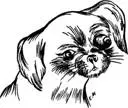
Though dislocated eyeball is not too uncommon in dogs generally, it is encountered most frequently in breeds with bulging eyeballs, such as the Boston Terrier and the Pekingese, because the combined factors of large eyeball and shallow eye socket render these dogs anatomically predisposed to eyeball dislocation.
Dislocation of the eyeball is always due to injury. Much less force is required to dislodge the eyeballs of Bostons or Pekingese than of the other breeds. In any case, the diagnosis of the condition is obvious, for the eyeball will be seen quite plainly to be dislodged from its socket to a variable degree. If the veterinarian is not immediately available, the eye should be washed continuously with a warm solution of boric acid made as described above. This is done in order to retard excessive dehydration of the eye tissues.
In the average case, the dislodged eyeball is easily replaced into its socket by gentle pressure and manipulation. The eyeball is then cleaned with boric-acid solution and flooded with boric-acid eye ointment. The eye is closed and covered with clean gauze over which is placed a pad of absorbent cotton; the eye is then bandaged snugly. The bandage serves to protect the injured eyeball and to secure it in its socket. The animal is now given a mild sedative such as an aspirin tablet. The eye is cleaned and the bandage replaced daily for two or three days. After this time, the bandage is removed altogether and boric-acid eye ointment is applied two or three times daily until no further evidence of eye inflammation is apparent.
Third eyelid and the gland of harder
On the inside corner of the eyeball is a membrane that is called the third eyelid or the nictitating membrane. This third eyelid acts as the windshield wiper of the eye by wiping away any foreign matter that may find its way to the surface of the eyeball. It can be seen rather easily when the animal repeatedly opens and closes its eyes.
Underneath this third eyelid is the Gland of Harder or, as it is commonly called, the haw. The Gland of Harder is never seen unless it is inflamed or tumefied.
When it is so affected, it appears as an unsightly, swollen protrusion at the inside corner of the lid. If the swelling does not recede after a couple of days of treatment with washings of boric-acid solution or applications of boric-acid eye ointment, it is advsisable to have the veterinarian remove it surgically. This is a rather simple operation.
An inflamed gland of harder
Paralysis of the third eyelid is quite common. In such cases, the membrane is plainly visible as a sheet of tissue, sometimes covering as much as half the front of the eyeball. It gives the dog an unsightly appearance and is uncomfortable to the animal. Treatment consists in surgical removal of the membrane by the veterinarian.